TUM
Klinikum rechts der Isar Technische Universität München
Neuro-Kopf-Zentrum
Abteilung für Neuroradiologie
Wissenschaftliche Auszeichnungen
Klinikum rechts der Isar Technische Universität München
Neuro-Kopf-Zentrum
Abteilung für Neuroradiologie
Wissenschaftliche Auszeichnungen
14:45 |
0147. 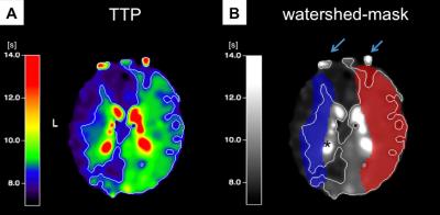
|
Analysis of cerebrovascular watershed areas in patients with high-grade carotid artery stenosis using DSC-based time-to-peak maps
Vanessa Griese, Stephan Kaczmarz, Anne Kluge, Kim van de Ven, Michael Helle, Hendrik Kooijman-Kurfuerst, Claus Zimmer, Christian Sorg , Jens Göttler, Christine Preibisch
Internal carotid artery stenosis (ICAS) is one of the leading causes for thromboembolic and hemodynamic cerebral infarction. Here, we present data from an ongoing clinical MRI-study in patients with asymptomatic, high-grade ICAS and healthy controls. Our major aim was to establish a method to delineate individual watershed areas in patients and controls using MRI-based dynamic susceptibility contrast (DSC) time-to-peak (TTP) maps, also including the anatomical information of magnetic resonance angiography (MRA) to define individual vascular territories. Watershed areas were enlarged and shifted in many of the vascular territories of stenosed carotid arteries, being verified by ss-pCASL in a subgroup.
|
14:33
|
0850. 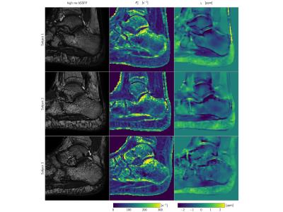
|
Simultaneous R2* and Quantitative Susceptibility Mapping of Trabecularized Yellow Bone Marrow: Initial Results in the Calcaneus
Maximilian Diefenbach, Jakob Meineke, Peter Foehr, Stefan Ruschke, Thomas Baum, Jan Kirschke, Andreas Hock, Hendrik Kooijman, Ernst Rummeny, Dimitrios Karampinos
R2* mapping has been previously used to measure trabecular bone density by quantifying the magnetic field inhomogeneity effects induced by the susceptibility difference between trabecular bone and marrow. Quantitative susceptibility mapping (QSM) has been emerging in other body parts for measuring the field-independent magnetic susceptibility. Trabecular bone is in many locations embedded in yellow fatty marrow. Therefore, trabecular bone QSM requires to account for the presence of fat. The purpose of the present work is to develop a methodology for simultaneous R2* mapping and QSM of trabecularized yellow bone marrow and apply the technique in the calcaneus of healthy volunteers.
|
 |
1108. |
27 |
Imaging of the Brachial Plexus using a 3D Dixon-TSE Pulse Sequence with Blood Vessel and CSF Signal Suppression: Preliminary Experience in Children
Barbara Cervantes, Amber Pokorney, Jan Kirschke, Patricia Cornejo, Jeffrey Miller, Dimitrios Karampinos, Houchun Hu
We demonstrate the clinical feasibility and utility of a 3D Dixon TSE sequence in imaging the brachial plexus nerves in children. The employed high spatial resolution MR Neurography (MRN) technique utilizes a refocusing flip angle train to maximize nerve signal and to suppress cerebrospinal fluid signal. Additionally, a T2-preparation with motion-sensitizing gradients was employed to suppress flowing blood signal and Dixon-based chemical-shift water-fat imaging was used to suppress fat signal in the head, neck, and chest. We illustrate this MRN technique in delineating the brachial plexus nerves and associated pathological conditions in 25 pediatric patients (age range: 6 months-21 years).
|